PHOENIX (AP) — Matt Hasselbeck took some nasty hits to the head during his 17-year NFL career, four years at Boston College and a childhood full of sports, but the quarterback seems to have emerged from more than two decades of collisions relatively unscathed, enjoying middle adulthood with his wife and three children.
The 50-year-old knows that some of his friends and former teammates haven't been as lucky.
That's why the three-time Pro Bowl selection recently spent two days at Boston University, going through a battery of scans and tests to determine how much all those collisions affected his brain. The hope is that it will be a small step toward a goal that has eluded doctors — diagnosing chronic traumatic encephalopathy in the living.
“They need guys like me — they need people who are feeling good to step up to the plate and volunteer,” Hasselbeck said. “I played quarterback, so I didn’t take some of the hits that a lot of my teammates took.
“But the guys who were blocking for me, or playing special teams, or trying to get the ball back for me on defense, I wanted to do something. They had my back, so in a way I’d like to have their back."
A degenerative brain disease that has been linked to concussions and other head trauma common in military combat and contact sports, CTE has been diagnosed in more than 100 former NFL players, casting a pall over the United States' most popular sports league. Some high-profile football players diagnosed with CTE after their deaths include Hall of Famers like linebacker Junior Seau and quarterback Ken Stabler, along with former Patriots tight end Aaron Hernandez.
Right now, CTE can only be diagnosed after death.
CTE can affect regions of the brain involved with regulating behavior and emotions. This can lead to memory loss, depression, violent mood swings and other cognitive and behavioral issues, though researchers note these symptoms can also be linked to other illnesses.
Hasselbeck's participation is just a small part of a study that Michael Alosco, co-director of clinical research at the Boston University CTE Center, hopes will expand to hundreds of participants in multiple locations. Alosco is the principal investigator in Boston and there are also sites in California, Arizona, Texas and Florida with potentially more to come.
The study seeks two types of people: Those who played professional or college football and those who have been diagnosed with Alzheimer’s disease and never played contact or collision sports and have no history of repetitive head impacts.
Alosco said there are many similarities between Alzheimer’s and CTE because both are degenerative brain diseases, but there are also some key differences, and mapping out those discrepancies is key to figuring out a way to diagnose CTE properly.
“We’ve applied what we’ve known worked for Alzheimer’s disease, but we’ve learned it does not work for CTE,” Alosco said. “We’ve really got to push discovery efforts and find the marker that’s specific to the protein of CTE versus Alzheimer’s because they’re different.
“How we’re going to get there is through the research we’re doing right now.”
Nicholas Ashton, the principal investigator in Phoenix, said roughly 10 patients have participated at the Arizona site. He said that currently, diagnosing CTE in the living is mostly guesswork because so many of the symptoms mimic Alzheimer's.
“The more people involved, the more information we get and the closer we get to an answer,” Ashton said. “Treatments exploded with Alzheimer's once we were able to accurately diagnose. We're trying to get to the same spot with CTE.”
Hasselbeck said he went through a variety of mental tests — like counting backward from 100 by sevens and reciting the alphabet backward. He also had a blood sample taken, an MRI on his brain and a PET (positron emission tomography) scan that injects a small amount of safe radioactive tracer that maps out the brain's function.
The former quarterback said the procedures were eye-opening and humbling.
“I've got a healthy respect for people who are dealing with serious brain trauma, whether it’s tumors or cancer,” Hasselbeck said. “I walked in there feeling completely healthy and you still get a little anxious when you’re doing a brain scan, an MRI. You come out and you’re looking at your brain.”
Hasselbeck also is donating his brain for research after he dies.
Not everyone in the study is a famous former NFL player.
Bryan Savage, a 56-year-old who lives in Maryland, is a former linebacker at Union College in Schenectady, New York — an NCAA Division III school. He traveled to Boston for the study and also has committed to donating his brain.
Savage said he's lucky to be relatively healthy but that he's been diagnosed with depression and ADHD over the past few years.
“I was interested because of some of the things that I've gone through physically and mentally,” Savage said. “You wonder if it's just getting older, a midlife crisis or if it's related to any brain trauma I might have had playing football.”
Savage praised the researchers who were with him during the study and said he was relieved to find out his brain scans look normal for a man in his mid-50s. He's reached out to some of his former teammates, encouraging them to get involved in the study.
“I'll talk to some of my former friends and they're going through a lot of the same things,” Savage said. “They'll say — ‘Why do I feel like this? I’ve never been depressed. Things that I used to be passionate about I don't care about anymore.'”
Alosco said the main reason Alzheimer's research is much more advanced than CTE research is sheer numbers: Alzheimer's research dates back decades and there have been countless people who have been studied.
By comparison, CTE research is relatively new. Volunteers like Hasselbeck and Savage are crucial to bridge the gap. The National Institutes of Health is sponsoring the current study, awarding $15 million for the research in 2025.
“The (Alzheimer's progress) is through years and years of research, of people donating their brains, and also collecting data during life,” Alosco said. “While we’ve been studying CTE for a while, we haven’t had the point where we’ve had all this data during life, that have also donated their brain. We need to get to that place.”
He added: “I’m really hopeful about detecting CTE — I don’t think that’s too distant. Once we can accurately diagnose, that’s when we can start some treatment trials.”
Hasselbeck — who played in the NFL for the Green Bay Packers, Seattle Seahawks, Tennessee Titans and Indianapolis Colts — said he had three concussions before he even started college, one playing football, another on the basketball court and another while goofing around while ice skating. He had a few concussions during his college or NFL career, but he wasn't sure of the exact number because some might have gone undetected since there was a culture of toughness in locker rooms, particularly early in his career.
Hasselbeck's pro career spanned from 1998 to 2015 and the quarterback said there was a huge change regarding head injuries during that time. Early in his career, the only acceptable answer after a hit to the head was “I'm good,” but by the mid 2010s, players were much more honest about potential concussions.
Hasselbeck's proud of that change. Now he hopes his participation in the current CTE study can continue that progress.
“I’ve always had a passion for making the game safer,” Hasselbeck said. “I love football. One of the things I’m most proud of as an NFL guy is not just as a player, but the entire time I worked with the league and NFLPA for ways to make the game safer.”
Former AP Sports Writer Jimmy Golen in Boston contributed to this report.
AP NFL: https://apnews.com/hub/NFL
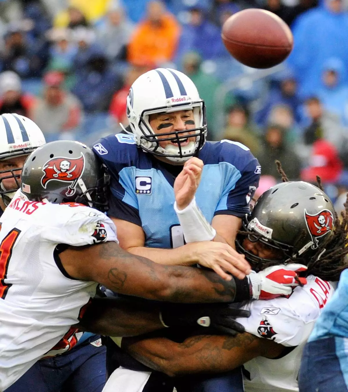
FILE - Tennessee Titans quarterback Matt Hasselbeck, center, throws as he is hit by Tampa Bay Buccaneers defenders Da'Quan Bowers, left, and Adrian Clayborn, right, in the first quarter of an NFL football game on Nov. 27, 2011, in Nashville, Tenn. (AP Photo/John Russell, File)
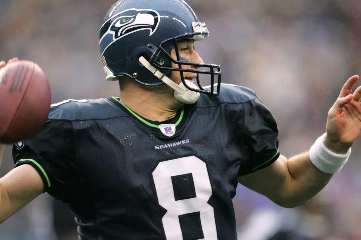
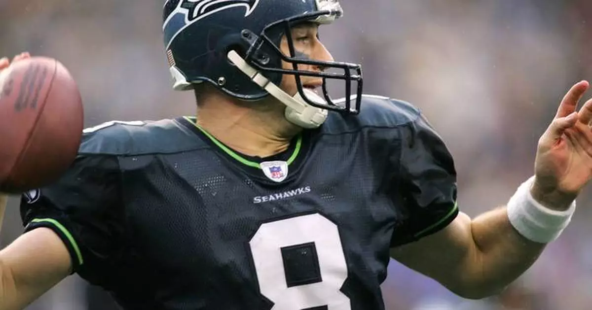
FILE - Seattle Seahawks quarterback Matt Hasselbeck passes during the fourth quarter against the Dallas Cowboys in Seattle, Oct. 23, 2005. (AP Photo/John Froschauer, File)
SAN FRANCISCO & PHILADELPHIA--(BUSINESS WIRE)--Aug 12, 2026--
Vivodyne, the company making human biology computable at AI scale, today announced the world’s largest human biological datacenter, with 12 robotic HIVE laboratories and the annual capacity to perform controlled trials on 3.1 million large human tissues per year — estimated at twice the scale of every clinical trial in the USA combined.
This press release features multimedia. View the full release here: https://www.businesswire.com/news/home/20260812148428/en/
The company also introduced its Series 2 TissueDisk, a wafer-scale biological chip that simultaneously grows hundreds of large, functional, living human tissues and is manufactured end-to-end on Vivodyne’s own robotic production line.
Eight of the world’s largest pharmaceutical companies have paid for early access to the platform. The goal? To discover and test new medicines ‘in humans’ before ever testing in people.
Together, the TissueDisk and Vivodyne’s robotic HIVE laboratories create something neither pharmaceutical research nor artificial intelligence has possessed before: a large-scale experimental environment in which the same reinforcement learning technique that has driven the explosion in AI language models can finally be harnessed to learn the workings of our physiology.
For pharmaceutical companies, that means learning how actual human tissue responds to a drug while decisions about targets, chemistry, dosing, and safety can still be made. Vivodyne’s approach allows millions of therapeutic interventions to be introduced into living human tissues, and their causal biological consequences measured directly with the most advanced, paired-data modalities available today: 3D scanning, transcriptomic sequencing, and deep proteomic analysis. It powers a self-driving experimentation loop, where an AI model can design massive experiments in human tissues, observe the real-world consequences, learn from the result, and optimize again and again, rather than working from static correlational data in published literature or computational predictions.
Vivodyne’s platform provides the foundation of the first world model of human biology. Previously, the controlled experiments required to reveal complex, physiological cause-and-effect could not be performed safely in patients or at nearly the needed scale. Vivodyne makes those experiments possible in living human tissue outside the body.
In this environment, the world is living human tissue. The intervention may be a drug, a novel combo regimen, or a gene therapy. The biological context may vary by donor or disease state. The consequences are measured across the tissue’s cells, blood vessels, immune components and molecular pathways.
“Superintelligence in biology is needed more than ever, because we’re running out of diseases that can be cured with the simple, single-target medicines of today,” said Andrei Georgescu, Ph.D., chief executive officer and co-founder of Vivodyne. “You cannot fix a car by turning a single screw, and the idea that the complex malfunctions in cancer, fibrosis, autoimmune disorders, or neurological disease can be fixed with a conventional single-target drug is wishful denial. We cannot keep hoping for medical miracles, and need to address the issue head-on: we do not understand human biology nearly well enough to predict exactly how to intervene. Testing in cells or animals is not enough; we don’t train our language models on whale sounds. To create AI that understands our intricate human biology, we need to continuously generate and train on huge amounts of human data, and we can’t get that by risking people. So we grow these functional human tissues by the millions instead; large, living tissues that grow their own blood vessels and immune cells and all the structures of native tissues. They mature, get diseases, bleed, scar, and, at huge scale, we learn how to make them heal. Every human response gives our AI something it cannot learn from a paper or a simulation: a living substrate to poke so that it can learn, from richer data than has ever been gathered, how it pokes back. At Vivodyne’s scale of automated human-tissue trials, all those learned consequences together become the training landscape for a world model of the human body, and the physical evidence that a pharmaceutical company needs before a drug is brought to patients.”
22 human organ systems, each a world of its own
Vivodyne grows over 20 types of different human organ tissues, both healthy and with patient-linked diseases, including liver, lungs, gut, bone marrow, pancreas, kidney, eyes, lymph nodes, and more, with disease-specific versions spanning fibrosis, site-specific solid tumors, inflammation, metabolic disorders, vascular disease, and countless others. The company trains causal, multimodal AI models on the experiments conducted within each organ type, alone and combined.
Connecting those models across organ systems builds a world model of the human body that can answer what happens when a pair of receptors is drugged, a biological pathway is interrupted, a therapy causes an unexpected side effect, how cells respond and communicate, and whether disease is aggravated, stopped, or reversed. These predictions can then be real-world tested at scale to confirm what actually happens in human tissue, and then refined and advanced.
“Computational models of human biology have largely been graded on how well they fill in data resembling information they have already seen, and that is reconstruction rather than novel prediction,” said Tony Bahinski, Ph.D., chief biotechnology officer of Vivodyne and long-standing member of the FDA’s science board. “Prediction means knowing what happens when you try something new. And in pharma, almost everything we try is new. Coding models don’t just memorize; they write and test enormous amounts of new code in their training, which is why they are so skilled today. So in kind, a biological world model can only truly learn by experimenting against complex, living human biology that can be dosed, perturbed, and allowed to recover. This training loop is what Vivodyne now makes possible at scale.”
Biological microprocessors, fully autonomous experiments
Each tissue on a Vivodyne TissueDisk functions like a biological microprocessor. Drug molecules and genetic perturbation are its input, and the living tissue computes the human response as an output.
Vivodyne manufactures TissueDisks on its own wafer-scale production line, enabling automated industrial scaling with the same exacting precision and reproducibility as semiconductor fabs.
TissueDisks run inside Vivodyne’s robotic HIVE laboratories. Each one operates automatically for weeks with full unattended automation, performing end-to-end cultivation, experimentation, and longitudinal data-gathering on tens of thousands of different tissues at a time, all containing billions of living cells. The automated labs grow tissues to maturity, deliver complex dosing regimens into their bloodstreams, perform multi-gene knockouts and activations, dose them with cell therapies or other complex modalities, capture timecourses of detailed three-dimensional scans and molecular or spatial analyses, running for weeks without human intervention.
Vivodyne’s software platform and scientific ontology, Hivemind, plans experiments, orchestrates its robotic laboratories, trains models on each result, and uses what it learns to determine which experiments should run next.
Each Vivodyne tissue starts with primary cells from human patients and grows to the size of the large medical biopsies studied by pathologists, each whole containing 200,000 to 500,000 cells of all the different types found natively. Those cells self-assemble into structures containing perfusable blood vessels, organ-specific function, and rich stroma and immune components. Because the vascular networks form through the same developmental process that produces blood vessels in the body, delivered test compounds arrive at the tissue through the bloodstream, just as they would in a patient.
This has resulted in unparalleled concordance with real human clinical results. For example, in direct comparisons to healthy and diseased patients, the cellular composition of Vivodyne airway tissue achieved a Lin’s concordance correlation coefficient of 96% with human patient airway tissue, with disease- and cell-specific gene expression indistinguishable from that seen in a typical sample of real patients.
The same system that generates training data for AI also answers the questions on which a drug program is built: it shows how new therapeutic targets causally respond in humans, which drug candidates or combinations produce the best responses, whether side-effect toxicities arise, and how donor-specific disease biology can change the outcome.
Vivodyne’s pharmaceutical programs span pulmonary tumors, multi-target biologics, cell therapies, immunosuppression, fibrosis, inflammatory disease, mRNA and lipid nanoparticles, chemotherapeutics, antibody-drug conjugates, vaccine development, and drug-induced liver injuries.
Partners have both reserved trial-scale experimental capacity within Vivodyne’s biological datacenters and increasingly work with Vivodyne on new drug-discovery programs.
“AI learned language from humanity’s entire body of written records,” Georgescu said. “It should learn medicine from the human body, too. Vivodyne is creating that human record of rich physiological data now, at the immense scale required to make human biology computable.”
Vivodyne is currently expanding data production from millions of tissues toward billions, and embedding their findings into an increasingly complete computational representation of the human body. Their mission is to restore and enhance human function.
About Vivodyne
Vivodyne makes human biology computable. The company trains medical superintelligence through robotic experimentation on millions of living, vascularized, functional human tissues that are lab-grown to the size of large clinical biopsies. Their technology produces human evidence before clinical trials, and powers AI-scale automated reinforcement learning environments where actions produce causal, clinically concordant consequences. Vivodyne’s human biological datacenters are located in the San Francisco Bay Area and Philadelphia. For more information, visit www.vivodyne.com.
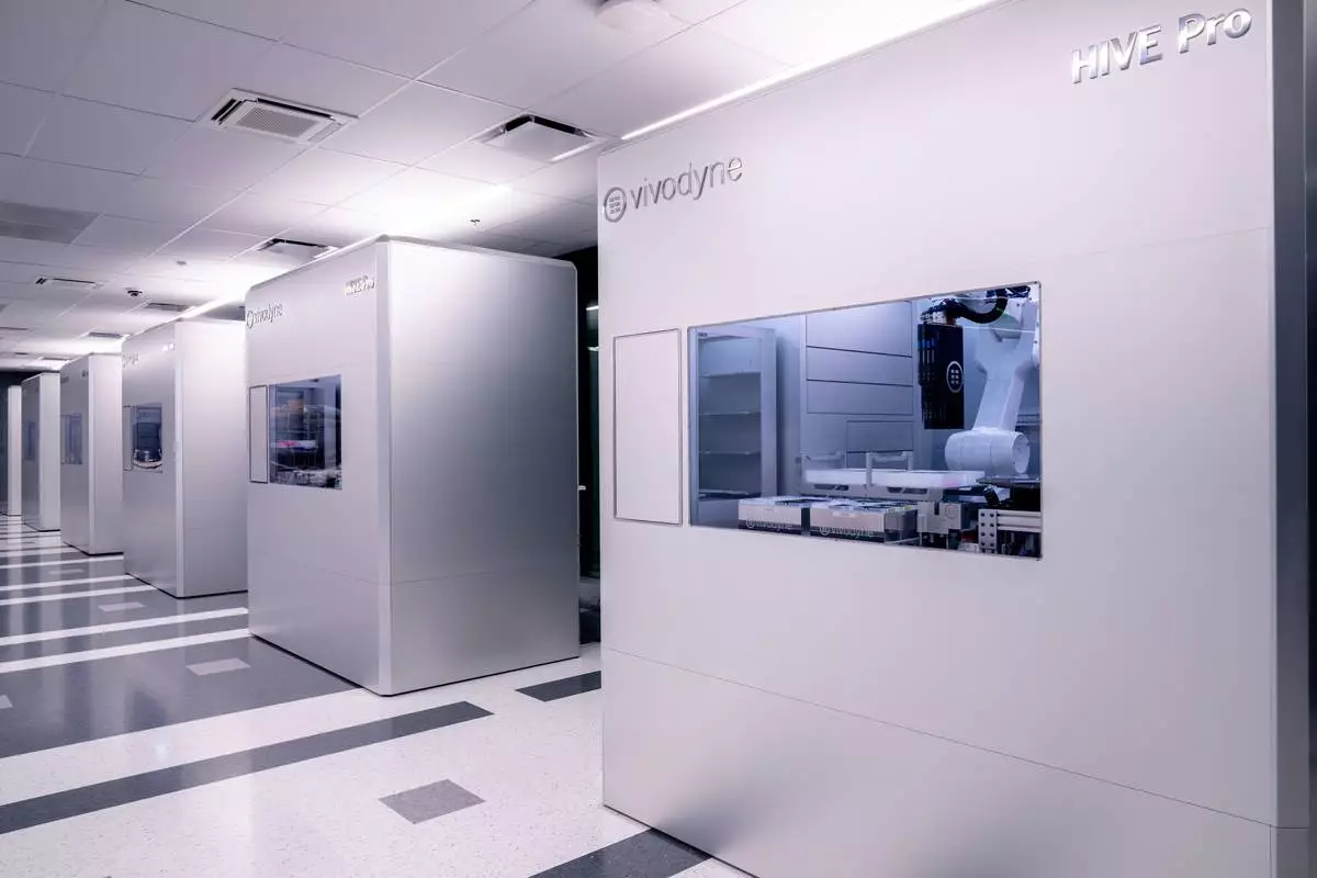
A row of automated human-tissue testing machines in Vivodyne's Human Datacenter.